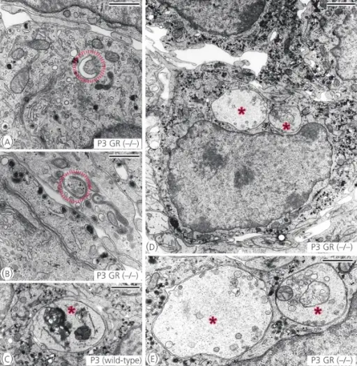
Electron microscopic characterisation of degenerated chromaffin cells located in the organ of Zuckerkandl (OZ) of postnatal day (P) 3 GRDBHCre mice (−/−) and corresponding wild-type littermates (+/+). (a, b) Showing an initial stage of autophagy, the formation of the isolation membrane (red circles). (c–e) Showing advanced stages of autophagy (red stars); cytoplasmic inclusions (autophagosomes) containing dense bodies (c), cytoplasm, vesicles and mitochondria [(d), enlarged in (e)]. Scale bars (a, b, e): 1 μm, (c, d): 2 μm.Cell loss and autophagy in the extra-adrenal chromaffin organ of Zuckerkandl are regulated by glucocorticoid signalling.
Schober A, Parlato R, Huber K, Kinscherf R, Hartleben B, Huber TB, Schütz G, Unsicker K - Journal of neuroendocrinology (2013). Not Altered. CC.
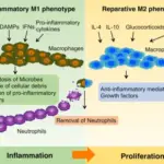
Autophagy is the natural, conserved degradation of the cell that removes unnecessary or dysfunctional components through lysosomal-dependent regulated mechanisms. Autophagy involves the formation of a double-membrane vesicle that encapsulates cytoplasm around targeted material for degradation that fuses with lysosomes for degradation.
Chaperone mediated autophagy is a proteolytic systems that contributes to degradation of intracellular proteins in lysosomes. Chaperone mediated autophagy substrate proteins are selectively targeted by ubiquitins and translocated into the lysosomal lumen through the coordinated action of chaperones located at both sides of the membrane and a dedicated protein translocation complex.