Axonal reaction are changes to axons that take place as a result of axonal damage. Axonal reaction most commonly indicates an axonal damage which follows damage to the cell body. The subsequent neuronal swelling is referred to as central chromatolysis.
What is Axonal Reaction?

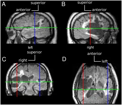
Case A1+ MRI FLAIR sequences.(A,B) Parasagittal sections through the left and right hemispheres. Left TG is atrophic and right TG is replaced by encephalomalacia (low signal intensity). Ischemic demyelination and retrograde degeneration within adjacent white matter regions appear as areas of high signal intensity. (C) Coronal section through the mid-portion of left and right TG and STG. (D) Horizontal section through left and right TG and STG. See text for image acquisition. Dissociation of detection and discrimination of pure tones following bilateral lesions of auditory cortex.
Dykstra AR, Koh CK, Braida LD, Tramo MJ - PloS one (2012). Not Altered. CC.


