Blastomyces fungal infection is a rare fungal infection usually acquired by breathing in the spores of the fungi blastomyces dermatitidis or blastomyces gilchristii.
What is the Pathology of Blastomyces Fungal Infection?
The pathology of blastomyces fungal infection is:
-Etiology: The cause of blastomyces fungal infection is inhaling airborne spores from contaminated soil into the lungs.
-Genes involved: Not applicable.
-Pathogenesis: The sequence of events that lead to blastomyces fungal infection occurs when aerosolized conidia are inhaled, and once in the lungs at body temperature, these conidia transform to the yeast phase. A self-limited infection develops in the majority of persons.
-Morphology: The morphology associated with blastomyces fungal infection shows small raised pus-filled (papulopustular) lesions. They may be violet colored and have very small abscesses around the borders of the lesions.
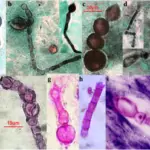
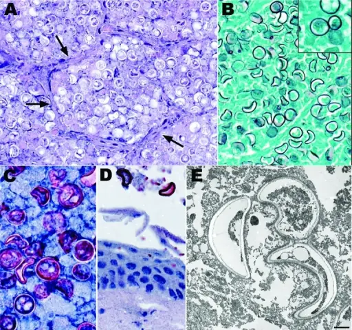
-Histology: The histology associated with blastomyces fungal infection shows marked pseudoepitheliomatous hyperplasia of epidermis, granulomatous and neutrophilic infiltrate, fungi are within giant cells.
How does Blastomyces Fungal Infection Present?
Patients with blastomyces fungal infection typically are all genders of age range 2 – 93 years. The symptoms, features, and clinical findings associated with blastomyces fungal infection include back pain, chest pain, fatigue, weight loss, poor appetite, shortness of breath, and fever.
How is Blastomyces Fungal Infection Diagnosed?
Blastomyces fungal infection is diagnosed by blood sample, urine sample, chest x-rays or CT scans of your lungs.
How is Blastomyces Fungal Infection Treated?
Blastomyces fungal infection is treated by antifungal medication Itraconazole.
What is the Prognosis of Blastomyces Fungal Infection?
The prognosis of blastomyces fungal infection is fair. Although some may recover completely, the mortality rate is about 30%.