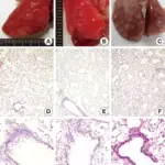
Cell injury is damage acquired to cells that may result in reversible or irreversible injury. Causes of cell injury may include physical agents, chemical agents, drugs, lack of oxygen, nutrient imbalances, infections, immune issues, or genetic issues. Cellular injury may involve morphologic alterations, reversible cell injury, or necrosis.
WHAT IS CELL INJURY?
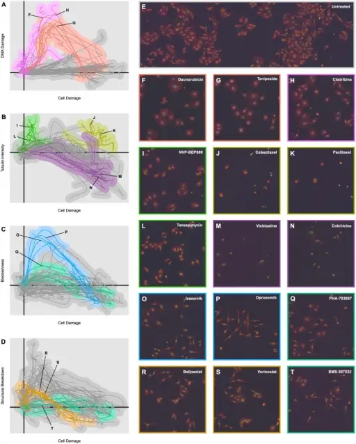
(A) Plot of the means of the phenotypic trajectories of all 154 compounds in two highly informative dimensions, “Cell Damage” and “DNA Damage”. Topoisomerase inhibitors (red) and antimetabolites (magenta) have been highlighted. (B) The plot of phenotypic trajectory means in the “Cell Damage” and “Tubulin Intensity” dimensions. Microtubule stabilizers (yellow), microtubule inhibitors (purple), and HSP90 inhibitors (green) have been highlighted. (C) The plot of phenotypic trajectory means in the “Cell Damage” and “Blebbishness” dimensions. Proteasome inhibitors (blue) and CDK inhibitors (teal) have been highlighted. (D) The plot of phenotypic trajectory means in the “Cell Damage” and “Structural Damage” dimensions. HDAC inhibitors (dark orange) and CDK inhibitors (teal) have been highlighted. (E) Example microscopy image of untreated cells. The red channel depicts α-tubulin intensity, the green channel Hoescht stain intensity, and the blue channel p-H2A.X stain intensity. (F–T) Example microscopy images of cells treated by 15 different compounds. Robust Classification of Small-Molecule Mechanism of Action Using a Minimalist High-Content Microscopy Screen and Multidimensional Phenotypic Trajectory Analysis: Twarog NR, Low JA, Currier DG, Miller G, Chen T, Shelat AA - PloS one (2016). Not altered. CC.
Post navigation
Previous Post
 What is Cellular Aging?