Cellular necrosis is identified as cell death due to multiple factors, such as cell swelling, loss of plasma membrane integrity, and ATP (adenosine triphosphate) depletion, which leads to calcium influx. Early research categorizes cell death in two distinct types: necrosis and apoptosis.
The morphology of necrosis microscopically is DNA degradation, hypertrophy of organelles, and the release of cytoplasmic content to provoke an inflammatory response. Under a microscope, the necrotic tissue may look like clumped cheese (crumbly and white). The patterns of tissue necrosis are based on the tissue and nature of the insult. They could be zonal, centrilobular, diffuse, multifocal, focal, and single-cell.
There are six characteristic morphologic patterns of necrosis which include:
- Fibrinoid necrosis
- Coagulative necrosis
- Liquefactive necrosis
- Gangrenous necrosis
- Caseous necrosis
- Fat necrosis
What is Fat Necrosis?
Fat necrosis is a type of necrosis of adipose tissue due to trauma or enzymatic activities that may result in saponification or calcification of adipose tissue. Sites that are prone to fat necrosis include the pancreas and breast tissue, although it can happen at other sites with adiposity.
What is Fibrinoid Necrosis?
Fibrinoid necrosis is a distinctive pattern of uncontrolled and irreversible cell death that appears as a result of antigen-antibody complexes. It is limited to the smaller blood vessels. Often the small arteries, glomeruli, and arterioles are impacted by an autoimmune disease (i.e., SLE or systemic lupus erythematosus). It is characterized by fibrinoid material.
What is Caseous Necrosis?
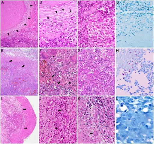
Caseous necrosis is a condition in which a walled off granuloma is formed. This is the classic type of necrosis caused in the lungs by tuberculosis. Grossly, it has a cheese like consistency.
What is Coagulative Necrosis?
Coagulative necrosis is a condition that happens from an infarct. It can emerge in every cell except for the brain. When observing a granuloma with central necrosis in the lungs of an individual with tuberculosis, note the Langhans-type giant cells. These cells can be observed in countless granuloma types and aren’t specific for tuberculosis.
What is Gangrenous Necrosis?
Gangrenous necrosis is the death of body tissue from insufficient circulation or grave bacterial infection.
What is Liquefactive Necrosis?
Liquefactive necrosis is distinguished by complete or partial transformation and dissolution of dead tissue into a viscous, liquid mass.


