Chiari type I malformation is an abnormality in which the cerebellum bulges through a normal opening in the skull where it joins the spinal canal. This puts pressure on parts of the brain and spinal cord, and may cause mild to severe symptoms. In most cases, the problem is congenital. There are several types of Chiari malformations, but type I is the most common.
What is the Pathology of Chiari Type I Malformation?
Etiology: The cause of Chiari Type I malformation is unknown.
Genes involved: Genes that have been implicated in Chiari type I malformations include: HOX gene, Noggin gene, EFNB1, TBX6, FGF2, PAX
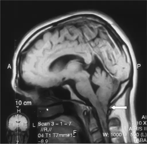
Pathogenesis: The sequence of events that lead to Chiari type 1 malformation are characterized by an inferior position of the cerebellar tonsils relative to foramen magnum. Some patients have an abnormal skull base or cervical segmentation abnormalities. Others have a small cranial vault or excess brain tissue.
Histology: The histology associated with Chiari type I malformation shows large and dysplastic fibrous tissue along with choroid plexus.
How does Chiari Type I Malformation Present?
Patients with Chiari type I malformation are more often females than males. The age range of the disease is mostly 25-45 years. The symptoms, features, and clinical findings associated with Chiari type 1 malformation include: sleep apnea, difficulty swallowing, muscle weakness, nystagus, headaches, and importantly the formation of a fluid filled pocket or cyst in the spinal cord, known as syrinx.
How is Chiari Type I Malformation Diagnosed?
Chiari type I malformation is diagnosed mostly by MRI. A CT scan can also be done.
How is Chiari Type I Malformation Treated?
Chiari type I malformation can be treated with painkillers if symptoms are present. A surgery is done to relieve pressure on the brain and restore the flow of CSF.
What is the Prognosis of Chiari Type I Malformation?
The prognosis of Chiari type I malformation is generally fair after surgery and depends on the extent of neurological deficit.


