Congestive splenomegaly can be due to chronic outflow obstruction. This may be due to obstruction of the splenic vein or extra hepatic portal vein. Congestive splenomegaly may lead to splenic or portal vein hypertension. Cirrhosis is a common cause of massive congestive splenomegaly. Schistosomiasis may also cause congestive splenomegaly.
WHAT IS CONGESTIVE SPLENOMEGALY?

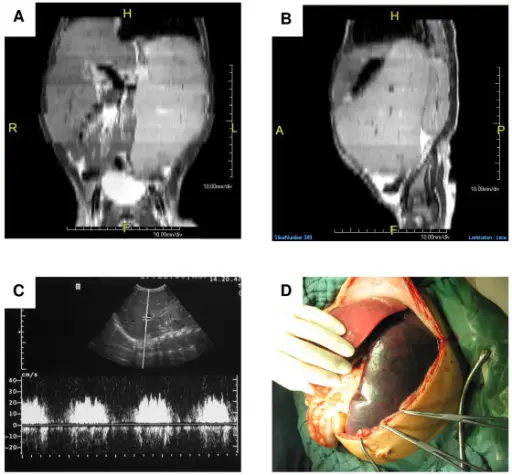
A) Magnetic resonance imaging (MRI) revealed enlargement of especially the spleen on axial projection. The splenic enlargement resulted in a compression of the bladder (H = Head, F = Foot, R = Right, L = Left). B) On sagittal MRI projection, compression of the left kidney was seen. The compression of other intraabdominal organs impeded alimentation (H = Head, F = Foot, A = Anterior, P = Posterior). C) On Doppler sonography, the portal vein was relatively prominent with a width of 1 cm. Portal vein blood flow was markedly increased. D) During surgery, the extent of splenomegaly was documented. Life-threatening hypersplenism due to idiopathic portal hypertension in early childhood: case report and review of the literature: Däbritz J, Worch J, Materna U, Koch B, Koehler G, Duck C, Frühwald MC, Foell D - BMC gastroenterology (2010). Not altered. CC.


