Cutaneous T-cell lymphoma aka mycosis fungoides is a cutaneous disorder characterized by localization of the neoplastic T- lymphocytes.
What is the Pathology of Mycosis Fungoides?
The pathology of mycosis fungoides is the scientific study of the pathology that arises from the presence of T-lymphocytes.
-Etiology: The cause of mycosis fungoides is unclear.
-Genes involved: None.
-Pathogenesis: The sequence of events that lead to mycosis fungoides involve Clonal growth of CD4 cells lacking the normal T cell antigens (CD7, CD5, CD 2).
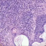
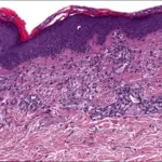
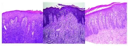
-Morphology: The morphology associated with mycosis fungoides shows lymphoid atypia in the skin.
-Histology: The histology associated with mycosis fungoides shows mixed perivascular infiltrate, dermal papillae fibrosis.
How does Mycosis Fungoides Present?
Patients with mycosis fungoides typically slightly high in females than males, present at an age range of over 50 years. The symptoms, features, and clinical findings associated with mycosis fungoides occurs in three forms Premycotic stage in which the lesions are erythematous, red-brown, scaly and pruritic, Infiltrative stage with slightly elevated, bluish-red, firm plaques and the Fungoid stage include pruritic plaques, itchiness, patches on the lower trunk and the gluteal region and ulceration.
How is Mycosis Fungoides Diagnosed?
Mycosis fungoides is diagnosed by physical exam and history taking, and skin biopsy.
How is Mycosis Fungoides Treated?
Mycosis fungoides is treated by topical medication, and chemotherapy.
What is the Prognosis of Mycosis Fungoides?
The prognosis of mycosis fungoides is good if the disease is not advanced.