Cysticercosis is a parasitic tissue infection caused by larval cysts of the tapeworm Taenia solium. These larval cysts infect brain, muscle, or other tissue, and are a major cause of adult onset seizures in most low-income countries.
What is the Pathology of Cysticercosis?
The pathology of cysticercosis is:
-Etiology: The cause of cysticercosis is larvae of the parasite Taenia solium
-Genes involved: Not applicable.
-Pathogenesis: The sequence of events that lead to cysticercosis are: after the ingestion of embryonated Taenia solium eggs, oncospheres hatch from the eggs, penetrate the intestinal wall into the circulation then penetrate the tissues of several organs neural or extra neural cells to form cysticerci in them.
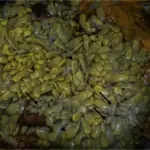
-Morphology: The morphology associated with cysticercosis shows fluid-filled cystic structures consisting of a thin bladder wall and parenchymatous portion containing a single invaginated scolex surrounded by a convoluted spiral canal. The hooks of the armed scolex may be visible in tissue sections.
-Histology: The histology associated with cysticercosis shows the cystic cavity contains the larval form: scolex with hooklets and two pairs of suckers.
How does Cysticercosis Present?
Patients with cysticercosis typically in children and young females aged 10-40 years. The symptoms, features, and clinical findings associated with cysticercosis include headaches, seizures, nausea, and vomiting.
How is Cysticercosis Diagnosed?
Cysticercosis is diagnosed by MRI, CT scans of the brain, blood tests to diagnose infection.
How is Cysticercosis Treated?
Cysticercosis is treated by antiparasitic therapy, corticosteroids, antiepileptic drugs, or surgery.
What is the Prognosis of Cysticercosis?
The prognosis of cysticercosis is good generally, however, it is mostly affected by the number of lesions and extent of inflammation. Individuals with one brain lesion have a very good chance of survival with no seizure relapses.