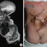
Encephalocele is a sac like protrusion of the brain and covering membranes through an opening in the skull. Encephalocele happens when the neural tube fails to close during pregnancy. Encephalocele can lead to the formation of a groove on the skull.
What is the Pathology of Encephalocele?
The pathology of encephalocele is:
Etiology: The cause of encephaloceles is the failure of the neural tube to close completely during fetal development. Both environmental and genetic factors have been seen to contribute to the cause of encephaloceles. Other causes include arsenic, tryptan blue, and teratogens.
Genes involved: Encephaloceles are more common in individuals who have a family history of neural tube defects such as spina bifida or anencephaly.
Pathogenesis: The sequence of events that lead to encephalocele is characterized by partial lacking of bone fusion. Hence, there is a gap through which a portion of the brain sticks out. Sometimes, the membranes that cover the brain along with the cerebrospinal fluid also protrudes out. The majority of encephaloceles are congenital.
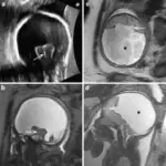
Histology: The histology associated with encephalocele shows dysplastic nerve tissue of cerebral and cerebellar cortex.
How does Encephalocele Present?
Patients with encephalocele typically present at age range of 1 day to 6 years old. Males and females are equally affected. The symptoms, features, and clinical findings associated with encephalocele include seizures, loss of balance, ataxia, and loss of strength.
How is Encephalocele Diagnosed?
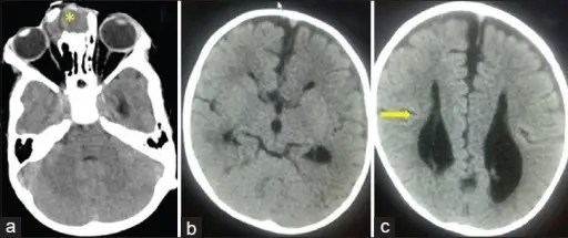
Encephalocele is diagnosed via prenatal ultrasound or physical exam. Other tests include fetal MRI, cell-free foetal DNA testing and amniocentesis.
How is Encephalocele Treated?
Encephalocele is treated with surgical intervention to place the protruding part of the brain and the membranes covering it back into the skull.
What is the Prognosis of Encephalocele?
The prognosis of encephalocele is poor if the patient develops complications.