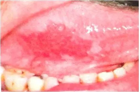
Erythroplakia is any lesion of the oral mucosa that presents as bright red velvety plaques which cannot be characterized clinically or pathologically as any other recognizable condition. Such lesions are usually irregular in outline, although clearly demarcated from the adjacent normal epithelium.
What is the Pathology of Erythroplakia?
The pathology of erythroplakia is:
-Etiology: The cause of erythroplakia is smoking.
-Genes involved: p53 gene.
-Pathogenesis: The sequence of events that lead to erythroplakia is epidermoid carcinoma (squamous cell carcinoma). Epidermoid carcinoma originates in abnormal mucosa as either leukoplakia, erythroplakia, or speckled leukoplakia. This disease most commonly begins in a leukoplakic lesion which can be smooth or rough, flat or elevated, ulcerated or intact.
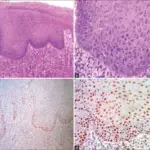
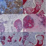
-Histology: The histology associated with erythroplakia reveals epithelial dysplasia, carcinoma in situ, and even invasive OSCC.
How does Erythroplakia Present?
Patients with erythroplakia typically affect males and females both present at the range of 25 and above. The symptoms, features, and clinical findings associated with erythroplakia include red macule or plaque with well-demarcated borders. The texture is characterized as soft and velvety. An adjacent area of leukoplakia may be found along with the erythroplakia.
How is Erythroplakia Diagnosed?
Erythroplakia is diagnosed only by biopsy.
How is Erythroplakia Treated?
Erythroplakia is treated by complete excision.
What is the Prognosis of Erythroplakia?
The prognosis of erythroplakia is poor.