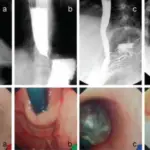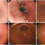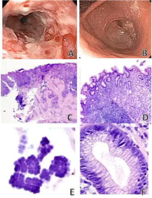
Brother’s endoscopic images. A) Severe erosive esophagitis in the distal third of esophagus. B) Marked narrowing (pinpoint lumen) of pylorus. Sister’s endoscopic images (four months later) were morphologically similar however there was complete pyloric obstruction secondary to peri-pyloric inflammation and edema. Histology in the siblings with co-existing Sarcina and H. pylori were nearly identical. C) Active ulcerating esophagitis (hematoxylin/eosin, ×200). D) Chronic active H. pylori gastritis (hematoxylin/eosin, ×400). E) Sarcina organisms in overlying gastric mucin showing characteristic basophilic tetrad packeted morphology (hematoxylin/eosin, ×600). F) H. pylori organisms in gastric pit (hematoxylin/eosin, ×400). H) pylori associated duodenitis was also present in both siblings (images not shown). Co-existence of Sarcina Organisms and Helicobacter pylori Gastritis/Duodenitis in Pediatric Siblings. Sauter JL, Nayar SK, Anders PD, D'Amico M, Butnor KJ, Wilcox RL - Journal of clinical & anatomic pathology (JCAP) (2013). Not Altered. CC.
Esophagitis is inflammation that may damage tissues of the esophagus, the muscular tube that delivers food from your mouth to your stomach. Esophagitis can cause painful, difficult swallowing and chest pain.
Examples of esophagitis include:
- Chemical esophagitis
- Eosinophilic esophagitis
- Infectious esophagitis
- Reflux esophagitis