Pathology of the female genital system is study of causes and effects of diseases related to the female reproductive system.
WHAT IS FEMALE GENITAL SYSTEM PATHOLOGY?

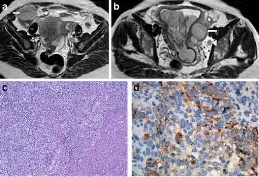
Endometrial tumour prolapsing into the cervical canal, without invasion of the cervical stroma and myometrium. The tumour extends bilaterally through the lumen of the tubes to the parametrial tissues (arrows). A left obturator lymph node is also seen (arrowhead). H&E section of the tumour with a solid pattern composed of large cells with pleomorphic nuclei, mitoses and scarce cytoplasm (c). These cells have small chromogranin-positive granules (d) and small synaptophysin granules in the cytoplasm (not shown) and are strongly positive for pan-cytokeratin CAM 5.2 (not shown). Neuroendocrine tumours of the female genital tract: a case-based imaging review with pathological correlation: Lopes Dias J, Cunha TM, Gomes FV, Callé C, Félix A - Insights into imaging (2015). Not altered. CC.


