Hemochromatosis or iron overload is a condition in which the body stores too much iron.
What is the Pathology of Hemochromatosis?
The pathology of hemochromatosis is:
-Etiology: The cause of hemochromatosis is the mutations in iron metabolism genes, such as HFE
-Genes involved: HFE.
-Pathogenesis: The sequence of events that lead to hemochromatosis involves iron to be absorbed by duodenal enterocytes via divalent metal transporter 1 (DMT1).
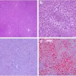
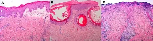
-Histology: The histology associated with hemochromatosis shows a pericanalicular pattern with iron appearing as a dark brown granular pigment, predominantly within hepatocytes.
How does Hemochromatosis Present?
Patients with hemochromatosis typically affect male present at age range of 40-60. The symptoms, features, and clinical findings associated with hemochromatosis include liver cirrhosis, diabetes mellitus, bronzing of the skin, joint pain, fatigue, and abdominal pain.
How is Hemochromatosis Diagnosed?
Hemochromatosis is diagnosed using liver biopsy, MRI, and blood tests.
How is Hemochromatosis Treated?
Hemochromatosis is treated with therapeutic phlebotomy/venesection or chelation therapy.
What is the Prognosis of Hemochromatosis?
The prognosis of hemochromatosis is good though, but poor after the development of cirrhosis, hepatocellular carcinoma, diabetes, or cardiomyopathy.