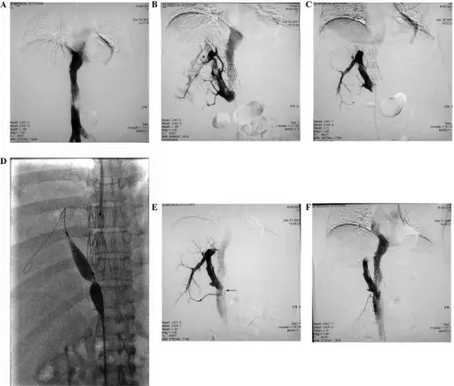
(A) Stent region shown by IVC angiography: The bloodstream was unobstructed within the stent, while stenosis occurred at the distal end of the vessel (indicated by the arrow). (B) In the AHV angiography performed through the femoral vein, a perforated membranous occlusion was shown at the opening of AHV, and a filling defect was generally observed in the vessel cavity. (C) Four days following the indwelling catheter thrombolytic therapy, the thrombus had predominantly dissolved, and only a small quantity of the embolus had shifted to the opening of AHV and blocked it (indicated by the arrow). (D) Four days following the thrombolytic therapy, a temporary filter was inserted through the jugular vein and a balloon catheter with a diameter of 10 mm was inserted through the femoral vein, by which the opening of the AHV was dilated. (E) Following the balloon dilation, the opening of the AHV remained narrow (indicated by the arrow). (F) Following the placement of a 12–40 mm stent, the vein cavity became unobstructed. Catheter-directed thrombolytic therapy combined with angioplasty for hepatic vein obstruction in Budd-Chiari syndrome complicated by thrombosis: Zhang Q, Xu H, Zu M, Gu Y, Wei N, Wang W, Gao Z, Shen B - Experimental and therapeutic medicine (2013). Not altered. CC.
Hepatic venous outflow obstruction is the condition where there is partial or complete obstruction of the hepatic veins.
Examples of hepatic venous outflow obstruction include:
- Hepatic vein thrombosis
- Sinusoidal obstruction syndrome
