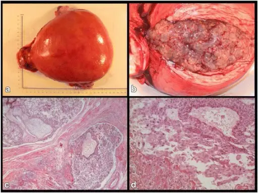
Hydatiform Mole. A photograph of the specimen with scale (cm); b Photograph of the specimen with longitudinal incision exposing vesicles; c Low magnification micrograph of the invasive complete hydatiform mole component (hematoxylin and eosin): two enlarged chorionic villi associated with a trophoblastic proliferation invading the vessels and the muscular wall of the uterus (bottom left); d high magnification micrograph of a suspected choriocarcinomatous component (hematoxylin and eosin): large syncitiotrophoblasts associated with a proliferation of atypical intermediate cytotrophoblasts. A HELLP syndrome complicates a gestational trophoblastic neoplasia in a perimenopausal woman: a case report. Vogin G, Golfier F, Hajri T, Leroux A, Weber B - BMC cancer (2016). Not Altered. CC.
Hydatiform mole is a rare complication of pregnancy characterized by the abnormal growth of trophoblasts, the cells that normally develop into the placenta. Trophoblastic tumor is also known as molar pregnancy.
Examples of hydatiform mole include:
- Complete mole
- Partial mole
- Invasive mole


