Hydrocephalus is the accumulation of excess fluid in the ventricles within the brain. Hydrocephalus can result in an increase in size of the ventricles. As a result, the pressure on the brain increases. Hydrocephalus iis due to excessive accumulation of cerebrospinal fluid within the ventricular system.
What is Hydrocephalus?

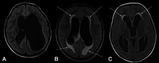
Axial FLAIR images of three different patients with hydrocephalus. In the first patient with chronic compensated hydrocephalus, lack of periventricular CSF resorption is seen (A). In the second patient, periventricular hyperintensity consistent with interstitial oedema due to acute decompansated hydrocephalus is demonstrated (arrow, B). Periventricular caps seen in middle aged adults should be differentiated from decompansated hydrocephalus (arrows, C). Evaluation of hydrocephalus and other cerebrospinal fluid disorders with MRI: An update.
Kartal MG, Algin O - Insights into imaging (2014). Not Altered. CC.


