The hyperacute rejection can occur in minutes of the transplant due to the presence of donor-specific antibodies. This is a very rare phenomenon and usually occurs in a patient with a history of transplantation. The CD4 T-cells react with HLA-class II molecules expressed by the cells of the transplant which initiates the immune response in the recipient and causes rejection of the organ within a few minutes of the transplant.
What is Hyperacute Rejection?

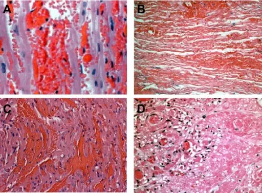
Hyperacute Rejection. Histopathology of xenograft rejection. The figure shows a comparison between anti-Gal and non-Gal antibody-mediated cardiac xenograft rejection. All panels show hematoxylin and eosin staining. A. Anti-Gal antibody-induced hyperacute rejection of a Gal-positive heart showing widespread intravascular hemorrhage characteristic of HAR. B. Anti-Gal antibody-mediated delayed xenograft rejection (DXR) of a Gal-positive heart on post-operative day 10. The rejected graft shows vascular injury, hemorrhage, and coagulative necrosis characteristic of anti-Gal-mediated DXR. C. Non-Gal antibody-mediated hyperacute rejection of a GTKO heart 90 min after reperfusion showing intravascular hemorrhage similar to that seen in Gal-mediated HAR (panel A). D. Non-Gal-mediated DXR on post-operative day 92 of a Gal-positive CD46 transgenic heart showing thrombotic microangiopathy. The recipient in panel D received chronic alpha-Gal polymer infusions to block anti-Gal antibody. Original magnification A and C 400×, B and D 200× (Panel C adapted from: McGregor CGA, et al. Cardiac xenotransplantation: progress toward the clinic. Transplantation. 2004: 78: 1569–1575.) Not Altered. CC.
Histopathologic insights into the mechanism of anti-non-Gal antibody-mediated pig cardiac xenograft rejection.
Byrne GW, Azimzadeh AM, Ezzelarab M, Tazelaar HD, Ekser B, Pierson RN, Robson SC, Cooper DK, McGregor CG

