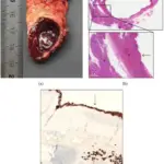
Hyposplenia is an anomaly characterized by the absence of spleen functionality tributary to a diversity of ailment states. Splenic tissues exist, however they are smaller than usual and non-functioning. Hyposplenia presents with imbalances in the two basic roles of the spleen; fighting infection and filtering of dysfunctional red cells.
What is Hyposplenia?
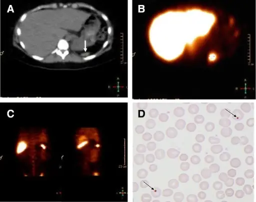
Hyposplenia. CT scan (A), spleen scintigraphy (B, C) and peripheral blood smear (D) of a woman with recurrent episodes of invasive infections withStreptococcus pneumoniae. A: CT scan showing horizontal section of a normal liver, while a very small rudimentary spleen is located at the tip of the arrow. B: Scintigraphy after injection of Technetium-99 m (99mTc) labeled, denatured red blood cells (RBC). The arrow shows uptake corresponding to the rudimentary spleen seen with the CT scan. C: Tc99m labeled, denatured RBC scintigraphy with different coronal sections of the abdomen showing the uptake corresponding to the rudimentary spleen seen with the CT scan. D: Peripheral blood smear stained with a standard H&E staining. Arrows point at red blood cells with Howell-Jolly bodies (HJB). Recurrent severe invasive pneumococcal disease in an adult with previously unknown hyposplenia. Ballegaard VC, Schejbel L, Hoffmann S, Kantsø B, Fischer CP - BMC infectious diseases (2015). Not Altered. CC.
Post navigation
Previous Post
 What is Asplenia?
Next Post
What are Accessory Spleens?