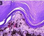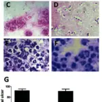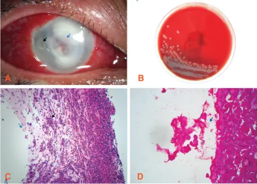
APseudomonasaeruginosa keratitis case. (A) Slit lamp microscopic image of severe central corneal infiltrate (blue arrow) with intensive conjunctival injection and a temporal satellite lesion (black arrow). Magnification: ×10. (B) Microbiological cultures obtained from a superficial corneal swab showed the presence of Pseudomonas aeruginosa. (C) Hematoxylin and eosin stains demonstrate that the corneal specimen contains numerous polymorphonuclear leukocytes (black arrow) and the epithelium and endothelium are absent (blue arrow). The lamellar architecture is lost and the frayed collagen is the result of widespread collagenolysis. Magnification: ×40. (D) Gram staining shows that Pseudomonas species could be found in the corneal deep stroma, which appear as short stubby rods and are Gram negative (blue arrow). Magnification: ×100.Pseudomonas aeruginosa keratitis misdiagnosed as fungal keratitis by in vivo confocal microscopy: a case report.
Hong J, Le Q, Deng SX, Cao W, Xu J - BMC research notes (2014). Not Altered. CC.
Keratitis is inflammation of the cornea caused by disease, injury, or infection. Contacts can cause keratitis by over wearing of contact lenses.
What is the Pathology of Keratitis?
The pathology of Keratitis is irritation of the cornea caused by a number of factors such as allergy, bacteria, fungi, viral infection and injury to the cornea.
How does Keratitis Present?
Keratitis presents with pain in the eye, and photophobia.
How is Keratitis Diagnosed?
Keratitis is diagnosed by histological examination, and medical history.
How is Keratitis Treated?
Keratitis is treated with antibiotics, eye drops, and surgery if indicated.
What is the Prognosis of Keratitis?
The prognosis of Keratitis gets better with medications and eradication of risk factors.