Mantle cell lymphoma is a lymphoproliferative disorder derived from a subset of naive pregerminal center cells localized in primary follicles or in the mantle region of secondary follicles.
What is the Pathology of Mantle Cell Lymphoma?
The pathology of mantle cell lymphoma is:
-Etiology: The cause of mantle cell lymphoma is not exactly known but approximately 85% of people with the condition have a genetic change, or mutation, in chromosomes 11 and 14.
-Genes involved: ATM, CCND1, TP53, MLL2, TRAF2 and NOTCH1 genes.
-Pathogenesis: The sequence of events that lead to mantle cell lymphoma starts from the chromosomal translocation t(11;14) resulting in aberrant expression of cyclin D1. Secondary genetic events increase the oncogenic potential of cyclin D1 and frequently inactivate DNA damage response pathways. In combination these changes drive cell-cycle progression and give rise to pronounced genetic instability.
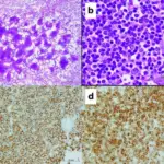
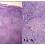
-Histology: The histology associated with mantle cell lymphoma shows expansion of the mantle zone that surrounds the lymph node germinal centers by small-to-medium atypical lymphocytes. These cells have irregular and indented nuclei, moderately coarse chromatin, and scant cytoplasm, resembling smaller cells of follicular lymphoma.
 Mantle Cell Lymphoma FISH. FISH image of MCL showing the CCND1 gene disruption (FISH, ×1,000); red/green signals marked the two ends of the CCND1 gene. A positive pattern showed two separate red and green signals. FISH, fluorescence in situ hybridization; MCL, mantle cell lymphoma. Clinicopathologic features of 112 cases with mantle cell lymphoma. Zhou DM, Chen G, Zheng XW, Zhu WF, Chen BZ - Cancer biology & medicine (2015). Not altered. CC.

How does Mantle Cell Lymphoma Present?
Patients with mantle cell lymphoma typically have a male-to-female ratio of 3:1 and present at age range of 35-85 years, with a median of 68 years. The symptoms, features, and clinical findings associated with mantle cell lymphoma include fever, night sweats, weight loss, generalized lymphadenopathy, abdominal distention from hepatosplenomegaly, fatigue from anemia or bulky disease.
How is Mantle Cell lymphoma Diagnosed?
Mantle cell lymphoma is diagnosed by lymph node biopsy and aspiration, bone marrow aspirate/biopsy, immunophenotyping and complete blood count (anemia, cytopenia and lymphocytosis). Body CT and PET scanning are important for initial staging.
How is Mantle Cell Lymphoma Treated?
Mantle cell lymphoma is treated by combination chemotherapy typically in combination with the monoclonal antibody rituximab (Rituxan), as first-line treatment, followed by autologous stem cell transplantation.
What is the Prognosis of Mantle Cell Lymphoma?
The prognosis of mantle cell lymphoma is poor. Mantle cell lymphoma is not curable with conventional chemoimmunotherapy. Overall, the median survival is approximately 6 to 7 years.