Mastocytosis is a disorder characterized by increased proliferation and accumulation of the mast cells in various organs of the body
What is the Pathology of Mastocytosis?
The pathology of mastocytosis is:
-Etiology: The cause of mastocytosis is the extreme activity of mast cells.
-Genes involved: KIT.
-Pathogenesis: The sequence of events that lead to mastocytosis, the consequence of the chronic and episodic release of mast cell mediators, and extreme buildup of mast cells in a tissue.
-Morphology: Rash.
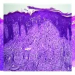
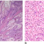
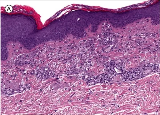
-Histology: The histology associated with mastocytosis shows infiltrates of the dermal mast cells around the blood vessels, round nuclei filled with cytoplasm.
How does Mastocytosis Present?
Patients with mastocytosis typically affect both males and females and the highest percentage are children and peaks at the age of 30-50 years. The symptoms, features, and clinical findings associated with mastocytosis include cutaneous lesions, flushing, headache, wheezing, diarrhea.
How is Mastocytosis Diagnosed?
The mastocytosis is diagnosed history taking and physical exam, CBC count, urinalysis, total tryptase level, bone scan and radiology.
How is Mastocytosis Treated?
The mastocytosis is treated by clearing the underlying symptoms such as the use of antihistamines, and topical corticosteroids.
What is the Prognosis of Mastocytosis?
The prognosis of mastocytosis is very good due to good response o the medication.