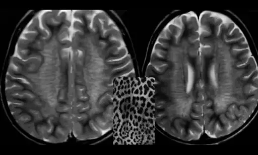
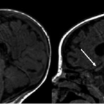
Metachromatic leukodystrophy is an inherited lysosomal storage diseases. Metachromatic leukodystrophy involves a progressive deterioration of motor and neurocognitive function.
What is the Pathology of Metachromatic Leukodystrophy?
Etiology: The cause of metachromatic leukodystrophy is deficient activity of arylsulfatase A, along with genetic disturbances.
Genes involved: Arylsulfatase A gene (ARSA gene), on chromosome 22q13.3.
Pathogenesis: Inability to degrade sulfated glycolipids, mainly the galactosyl-3-sulfate ceramides.
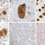
Histology: The histology associated with metachromatic leukodystrophy shows metachromatic granules. In the nervous system, the loss of myelinated oligodendrocytes is seen.
How does Metachromatic Leukodystrophy Present?
Patients with metachromatic leukodystrophy are on average 4 years old. Some cases are in the age range 12-14 years. The disease has equal occurrence in both sexes. The symptoms, features, and clinical findings associated with metachromatic leukodystrophy include loss of the ability to detect sensations, hearing loss, seizures, and psychosis.
How is Metachromatic Leukodystrophy Diagnosed?
Metachromatic leukodystrophy is diagnosed by laboratory testing.
How is Metachromatic Leukodystrophy Treated?
Metachromatic leukodystrophy currently has no treatment. Symptomatic supportive care is needed to address constipation, seizures, dystonias, spasticity, and difficulty feeding.
What is the Prognosis of Metachromatic Leukodystrophy?
The prognosis of metachromatic leukodystrophy is poor. Most children within the infantile form die before 6 years old.


