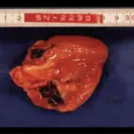
Microscopic polyangiitis is another form of necrotizing vasculitis that generally affects capillaries, as well as small arterioles and venules.
What is the Pathology of Microscopic Polyangiitis?
The pathology of microscopic polyangiitis is:
-Etiology: The cause of microscopic polyangiitis is not well understood but there is the involvement of the inflammatory response.
-Genes involved: Unknown.
-Pathogenesis: The sequence of events that lead to microscopic polyangiitis is not well known but there is the involvement of ANCA which is an autoimmune response
-Morphology: NA.
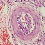
-Histology: The histology associated with microscopic polyangiitis shows segmental fibrinoid necrosis, focal transmural necrotizing lesions; granulomatous inflammation is absent.
How does Microscopic Polyangiitis Present?
Patients with microscopic polyangiitis typically are males that present at the age range of 50 years old. The symptoms, features, and clinical findings associated with microscopic polyangiitis include hemoptysis, hematuria, proteinuria, abdominal pain or bleeding, muscle pain or weakness, and palpable cutaneous purpura.
How is Microscopic Polyangiitis Diagnosed?
Microscopic polyangiitis is diagnosed by physical examination, renal function tests, chest radiography, GIT endoscopy.
How is Microscopic Polyangiitis Treated?
Microscopic polyangiitis is treated with corticosteroids, immunosuppressors, and intravenous immunoglobulin.
What is the Prognosis of Microscopic Polyangiitis?
The prognosis of microscopic polyangiitis is good since it has a reasonable survival rate of 90%.