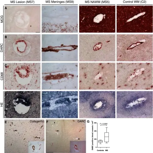
DARC expression in human subcortical white matter of control and multiple sclerosis cases. Immunohistochemical analysis of brain vessels within the human cortex from multiple sclerosis (MS) cases with active inflammatory demyelinating lesion (first column, MS lesions), within meninges (second column, MS Meninges), within normal appearing white matter (third column, MS NAWM) and from a control case within subcortical white matter (fourth column, Control WM) are shown. MOG: MOG immunohistochemistry for myelin differentiating demyelinating lesions from normal myelinated areas in cerebral cortex and subcortical cortical white matter in multiple sclerosis and controls (A). DARC: DARC-positive blood vessels were detected in all areas in multiple sclerosis cases and in controls (B). CD68: CD68 immunohistochemistry detects inflamed blood vessels (first panel) as well as activated microglia throughout the white matter (C). HE: Histology shown by haematoxylin and eosin staining of the corresponding area (D). Scale bar = 50 μm. Bottom: Collagen IV immunohistochemistry in multiple sclerosis normal appearing white matter identifying all blood vessels (E), whereas DARC staining could only be detected in a subset of blood vessels (F, arrows). DARC immunoreactivity was detected in larger (large arrow, higher magnification in inset) as well as in smaller blood vessels (small arrow). Insets show area around the blood vessel highlighted by the large arrow. Arrowhead points to a blood vessel, which is DARC-negative. Scale bar = 500 μm. (G) Quantification showing more DARC-positive blood vessels in multiple sclerosis normal appearing white matter compared with control white matter (P-value = 0.0282).DARC shuttles inflammatory chemokines across the blood-brain barrier during autoimmune central nervous system inflammation.
Minten C, Alt C, Gentner M, Frei E, Deutsch U, Lyck R, Schaeren-Wiemers N, Rot A, Engelhardt B - Brain : a journal of neurology (2014). Not Altered. CC.
Multiple sclerosis is autoimmune inflammatory disease that attacks myelinated axons.
What is the Pathology of Multiple Sclerosis?
Etiology: The cause of multiple sclerosis is unknown.
Genes involved: HLA region of chromosome 6. Changes in this area increase the probability of getting MS.
Pathogenesis: The sequence of events that lead to multiple sclerosis are the formation of lesions in the central nervous system (also called plaques) on myelin due to autoimmune attack.
Histology: The histology associated with multiple sclerosis shows multiple focal areas of myelin loss within the CNS called plaques or lesions, macrophages, and gliosis.
How does Multiple Sclerosis Present?
Patients with multiple sclerosis are most common in the age range 20-40 years. Females are more commonly affected. The symptoms, features, and clinical findings associated with multiple sclerosis include dizziness, fatigue, movement issues, and eye issues.
How is Multiple Sclerosis Diagnosed?
Multiple sclerosis is diagnosed primarily by ruling out other conditions and looking at the history and clinical findings. Tests include evoked potential, laboratory tests, spinal tap, and MRI.
How is Multiple Sclerosis Treated?
Multiple sclerosis has no cure, and is symptomatically managed.
What is the Prognosis of Multiple Sclerosis?
The prognosis of multiple sclerosis is good. Most people with multiple sclerosis have a relapsing-remitting disease course.