Myelomeningocele is a type of spina bifida, in which the bones of the spine do not completely form. This results in an incomplete spinal canal through which the meninges and spinal cord protrude from the back.
What is the Pathology of Myelomeningocele?
Etiology: The causes of myelomeningocele include environmental, maternal and genetic factors.
Genes involved: Some genetic factors are involved such as the presence of chromosomal anomalies of trisomy 18 or 13. Family history of an affected twin or first-degree relative is also important.
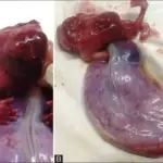
Pathogenesis: The pathology of myelomeningocele is characterized by the incomplete closure of the spinal neural tube during the first month of pregnancy. Hence, the neural tissue and meninges protrude outwards. This exposed neural tissue is called a placode. The existence of a placode with the meningeal lining is a hallmark of myelomeningocele. Hence, the meninges along with the neural tissue protrude outwards. The spinal cord herniates outwards in the fluid filled sac.
Histology: The histology associated with myelomeningocele shows elements of all germ layers. The squamous epithelium or fibrous connective tissue covers the dome of the myelomeningocele that comprises of the neural placode. There may also be the presence of meninges, blood vessels, and hyalinized connective tissue.
How does Meningomyocele Present?
Patients with meningomyocele comprise of more females than males. Meningomyocele is usually obvious at birth.
How is Myelomeningocele Diagnosed?
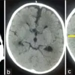
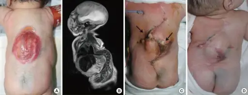
Myelomeningocele is diagnosed by physical exam. Earlier diagnostic methods include ultrasound, and maternal serum alpha fetoprotein test.
How is Myelomeningocele Treated?
Myelomeningocele is treated by surgery.
What is the Prognosis of Myelomeningocele?
The prognosis of myelomeningocele is fairly good, and lifespan is generally normal.