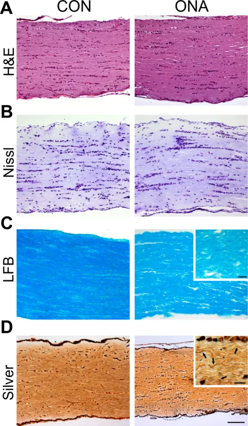
Optic nerve pathology in optic nerve homogenate antigen animals. A: Longitudinal optic nerve sections from both groups (control [CON] and optic nerve homogenate antigen [ONA]) stained with hematoxylin and eosin (H&E) showed no inflammatory cells (n=8/group). B: Optic nerve sections were also stained with Nissl. In neither group were cellular abnormalities noted, but the axons of ONA nerves were more disorganized (n=8/group). C: Luxol fast blue (LFB) staining was applied to assess signs of demyelination (n=8/group). Particularly in the magnified image, it can be clearly noted that axons of the ONA group are more disorganized, but still appear to be myelinated. D: Nerves were stained with Bielschowsky silver impregnation for axon evaluation (n=8/group). In ONA sections, many more damaged or swollen axons were noted. In the magnified section, arrows indicate swollen axons and arrowheads injured axons (scale bar 100 µm and higher magnification 10 µm).Immune response against ocular tissues after immunization with optic nerve antigens in a model of autoimmune glaucoma.
Joachim SC, Reinehr S, Kuehn S, Laspas P, Gramlich OW, Kuehn M, Tischoff I, von Pein HD, Dick HB, Grus FH - Molecular vision (2013). Not Altered. CC.
Optic nerve pathology deals with the spectrum of diseases targeted towards the nerve of vision.
Examples of optic nerve pathology include:
- Anterior ischemic optic neuropathy
- Papilledema
- Optic nerve damage
- Optic neuritis
- Other optic neuropathies
 What is Retinal Lymphoma?