Pancreas divisum is a fusional anomaly of the ductal systems of the dorsal and ventral buds of the embryonic pancreas during the seventh week of intrauterine life.
What is the Pathology of Pancreas Divisum?
The pathology of pancreas divisum is:
-Etiology: The cause of pancreas divisum is congenital fusion failure of the dorsal and ventral buds ducts.
-Genes involved: CFTR gene.
-Pathogenesis: The sequence of events that lead to pancreas divisum caused a failure of the ducts of the dorsal and ventral buds to fuse throughout embryologic growth, at around the 8th week of intrauterine life.
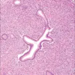
-Morphology: None.
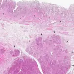
-Histology: No difference between normal pancreatic tissue, acini and ductules.
How does Pancreas Divisum Present?
Patients with pancreas divisum typically slightly high in females present at age range of between infancy and adulthood. The symptoms, features, and clinical findings associated with pancreas divisum include epigastric tenderness. palpable pseudocyst, abdominal pain, and pancreatitis.
How is Pancreas Divisum Diagnosed?
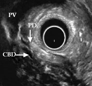
Pancreas divisum is diagnosed through radiological study- endoscopic retrograde cholangiopancreatography (ERCP), CT scan, magnetic resonance cholangiopancreatography (MRCP), and clinical presentations.
How is Pancreas Divisum Treated?
Pancreas divisum is treated through surgical intervention sphincteroplasty, and minor papilla sphincterotomy. Medical management of symptoms may be utilized.
What is the Prognosis of Pancreas Divisum?
The prognosis of pancreas divisum is good with appropriate management.