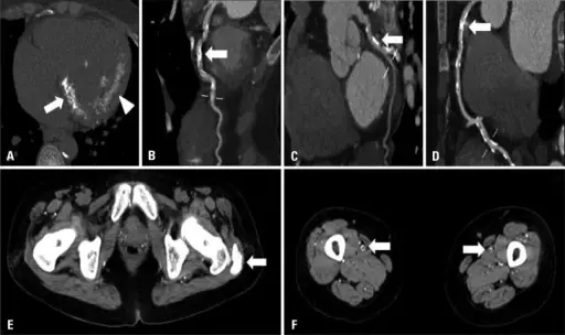
Cardiac CT (A-D) and peripheral CT (E and F) show extensive calcification. Cardiac CT shows severe mitral valve calcification (arrow) and myocardial calcification (arrowhead, A), diffuse calcification of LAD coronary artery (B), LCX coronary artery (C), and RCA coronary artery (D). Non-enhanced CT shows muscular calcification (arrow, E) and superficial femoral artery calcification (arrows, F). Porcelain heart: rapid progression of cardiac calcification in a patient with hemodialysis: Lee HU, Youn HJ, Shim BJ, Lee SJ, Park MY, Jeong JU, Gu GM, Jeon HK, Lee JE, Kwon BJ - Journal of cardiovascular ultrasound (2012). Not altered. CC.
Pathologic calcification is calcification at sites that are not normally calcified. The two types include dystrophic calcification and metastatic calcification.
Dystrophic Calcification
Metastatic Calcification
