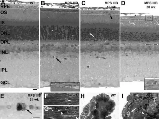
Eye Pathology.Light micrographs showing pathology from normal (A, F, H) and MPS IIIB (B–D, E, G, I) mouse retinas (A–E), sclera (F, G), and ciliary bodies (H, I). (A) Normal histology from a 4-week old wild type mouse retina is shown and is indistinguishable from older wild type retinas at 45 weeks. (B) In the 8-week old MPS IIIB mouse retina, aberrant lysosomal storage can be seen in non-neuronal cells in the inner retina (arrow). Inset: Localized disruption of the retinal pigment epithelium (RPE) from a nearby region of the same retinal section is shown. (C) At about 16 weeks, the outer segments (OS) of the MPS IIIB mouse are shortened, the outer nuclear layer (ONL) is reduced by 2–4 rows of nuclei, and pyknotic nuclei are seen in the ONL (arrow). Macrophage-like cells are present in the subretinal space (arrowhead). (D) In the 30-week old MPS IIIB mouse retina, OSs are further shortened and the ONL is reduced by nearly half. Inset: higher magnification of the boxed area showing dense, round melanosome-like structures in the RPE. (E) By 34 weeks, a subpopulation of cells in the ganglion cell layer (GCL) has a dense appearance with numerous lysosomal inclusions in the cytoplasm. (F) Normal histology from a 4-week old wild type mouse sclera is shown. (G) Lysosomal storage appears as pale vesicles in the sclera of the MPS IIIB mouse (arrowheads). (H) Normal histology from a 4-week old wild type mouse ciliary body is shown. (I) By 30 weeks of age, the ciliary body of the MPS IIIB mouse appears disorganized and swollen with lysosomal storage vesicles (arrow). Numerous dense, round melanosome-like structures are seen (arrowhead). IS, photoreceptor inner segments; OPL, outer plexiform layer; INL inner nuclear layer; IPL, inner plexiform layer. Smaller bar = 10 micron for A–D, H, I. Larger bar = 10 micron for E–G.Development of sensory, motor and behavioral deficits in the murine model of Sanfilippo syndrome type B. Heldermon CD, Hennig AK, Ohlemiller KK, Ogilvie JM, Herzog ED, Breidenbach A, Vogler C, Wozniak DF, Sands MS - PloS one (2007). Not Altered. CC.
Pathology of the sclera deals with the varieties of disorder that alters the proper function of the sclera.
Pathology of the sclera may include:
- Necrotizing scleritis due to rheumatoid arthritis.
