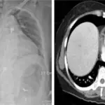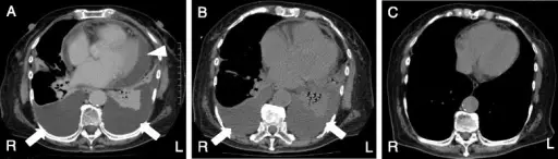
Pericardial effusion is an abnormal fluid accumulation in the pericardial space.
What is the Pathology of Pericardial Effusion?
Pericardial effusion pathology is characterized by fluid buildup in the pericardial space that may be serious with fibrin strands, serosanguineous fluid, blood, pus, or chyle, that may eventually lead to cardiac tamponade.
How does Pericardial Effusion Present?
Pericardial effusion presents as chest discomfort, light-headedness, palpitations, cough, dyspnea, hoarseness and hiccups.
How is Pericardial Effusion Diagnosed?
Pericardial effusion diagnosis includes echocardiography and electrocardiogram.
How is Pericardial Effusion Treated?
Pericardial effusion treatment includes pericardiocentesis, pericardiostomy, and use of aspirin, colchicine, steroids, and antibiotics.
What is the Prognosis of Pericardial Effusion?
Pericardial effusion prognosis is good, however poor prognosis is attributed to fever >38C, traumatic cause, unresponsive to NSAIDs, and in patients on anticoagulants.