Peritoneal cavity Infections is a silk-like membrane that lines your inner abdominal wall and covers the organs within your abdomen that is usually due to a bacterial or fungal infection.
What is Peritoneal Cavity Infections?

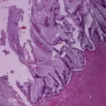
Schematic representation of liver fibrosis and cirrhosis associated with fascioliasis.(A) Normal architecture of a sheep's liver. Haematoxylin and eosin (HE) staining. Taken from Ref. [19]. (B) Juvenile parasite migrating in the peritoneal cavity causing destruction and haemorrhages in case (HE stain, magnification x100). The biopsy in the peritoneum was performed due to clinical suspicion of metastases Taken from Ref. [48]. (C) Typical microscopic appearance (400X) of liver from the infected sheep at 8 weeks post infection, massive infiltration of inflammatory cells and deposition of collagen can be observed. Taken from Ref. [19]. (D) Liver cirrhosis caused by F. hepatica infection. Trichrome stain (100x). Rat. Authors’ photo gallery. (E) Extensive fibrosis (Fi) with formation of fibrotic nodules (FN), architectural disruption of the liver and regeneration; stage IV or F4 (cirrhosis). Trichrome stain. Rats. Authors’ photo gallery. Association of Fasciola hepatica Infection with Liver Fibrosis, Cirrhosis, and Cancer: A Systematic Review
PLoS Neglected Tropical Diseases. Not Altered. CC.
Post navigation
Previous Post
 What are Tumors of the Appendix?
Next Post
What is Sclerosing Retroperitonitis? 