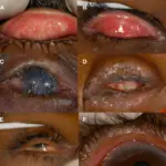
Pinguecula is an abnormal growth of the tissue on the conjunctiva. There may be a yellow limbus associated with submucosal elevation on the conjunctiva due to actinic damage.
What is Pinguecula?
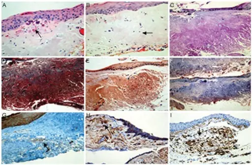
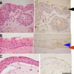
Immunohistochemical staining of pinguecula autofluorescence (AF). (A) Conjunctiva stroma with thinning of the overlying epithelium and calcification were detected. (B) Eosinophilic-stained amorphous material was detected in the subepithelial layer of the conjunctiva. (C-F) No immunoreactivity was detected. (G) Immunoreactivity to transglutaminase-2 was detected in the normal epithelium, normal vessel walls, and elastotic degeneration area of AF. (H,I) Immunoreactivity was detected. (A,B) Hematoxylin-eosin, (C) periodic acid-Schiff, (D) Masson's trichrome, (E) Congo red, (F) Oil Red O, (G)
transglutaminase-2, (H) CD29 (β-1-integrin), and (I) CD 34 (×400). Not Altered. CC.
Post navigation
Previous Post
 What is Conjunctival Scarring?
Next Post
What is Pterygium?