Proptosis is the bulging of the eyes causing a positional shift of the eyes in the socket.
What is Proptosis?
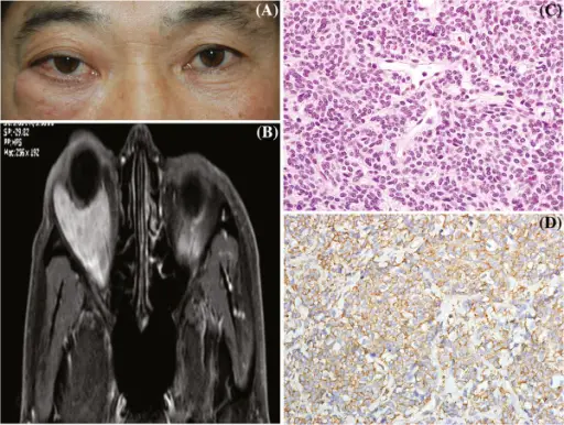
Photographs demonstrating right lower lid swelling and proptosis of the right eye (A). T1-weighted magnetic resonance imaging demonstrated a large, lobulated, smooth-margined mass in the lower lateral aspect of the right orbit, measuring 28x33x32mm in volume with a low signal that diffusely enhanced with gadolinium (B). Tumor cells were arranged in organoid and sheet-like patterns, and contained intimately associated capillary-sized blood vessels. Individual tumor cells were round, oval, polyhedral, or fusiform. Nuclei were round or ovoid and centrally placed in pale or slightly eosinophilic cytoplasm. Cell borders were not clearly delineated. Tumor cells were intermingled with thin fascicles of bland spindle cells (hematoxylin-eosin, original magnification x400) (C) and showed strong cytoplasmic positivity for smooth muscle actin (SMA) (immunoperoxidase/hematoxylin counterstain, original magnification x400) (D).Orbital glomus tumor in an Asian patient.
Chang M, Lee Y, Baek S, Lee TS - BMC ophthalmology (2012). Not Altered. CC.
Post navigation
Previous Post
 What are other Inflammatory Conditions of the Orbit?
Next Post
What is Wegener Granulomatosis?