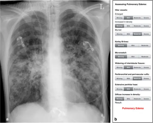
Pulmonary edema is a life-threatening emergency characterized by excessive interstitial fluid that accumulates in the alveolar space.
What is the Pathology of Pulmonary Edema?
The pathology of pulmonary edema is: The anomaly resulting from hemodynamic disturbances (hemodynamic or cardiogenic pulmonary edema) or direct increases in capillary permeability, owing to microvascular injury.
-Etiology: The cause of pulmonary edema is, lymphatic impediment, injury to the alveolar-capillary barrier, reduced plasma oncotic pressure, augmented negative interstitial pressure, and Idiopathic mechanism.
-Genes involved: None.
-Pathogenesis: The sequence of events that lead to pulmonary edema is as follows, Augmented pressure/pooling, leads to amplified pulmonary venous pressure, which leads to augmented pulmonary capillary pressure further leading to fluid in interstitial spaces causing augmented pressure in Interstitial spaces hence resulting to pulmonary edema (fluid in alveoli).
-Histology: The histology associated with pulmonary edema shows the pulmonary venules and alveolar capillaries are dilated and engorged with blood. The alveolar walls are thickened due to edema and later due to fibrosis. The alveolar spaces contain RBCs and many large macrophages with brownish hemosiderin pigment granules.
How does Pulmonary Edema Present?
Patients with pulmonary edema typically Men are affected more than women, present at age range of above 50 years. The symptoms, features, and clinical findings associated with pulmonary edema include breathlessness, wheezing, anxiety, sweating, frothy, blood-tinged sputum, and tachypnoeic with peripheral shutdown
How is Pulmonary Edema Diagnosed?
Pulmonary edema is diagnosed in Laboratory studies, complete blood count to assess anemia. pulse oximetry assessing hypoxia, arterial blood gas analysis, blood urea nitrogen level. Imaging studies, electrocardiography, CT scanning.
How is Pulmonary Edema Treated?
Pulmonary edema is treated as an emergency. General, Addressing (ABC) Airway, Breathing, Circulation. Prop up, oxygen therapy, mechanical and ventilatory support, IV access, and monitoring urine output. Underlying cause treatment, Noninvasive pressure-support ventilation CPAP, and BiPAP. Nitroglycerin for preload reduction. Diuretics. ACE inhibitors for afterload reduction.
What is the Prognosis of Pulmonary Edema?
The prognosis of pulmonary edema is poor especially in patients with congestive heart failure.