Retinal detachment is separation of the neurosensory retina from the retinal pigment epithelium.
What is Retinal Detachment?
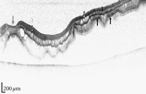
Horizontal spectralis OCT of the retinal detachment shown in Figure 1, scan direction is indicated by the white dotted line in Figure 1. Characteristic changes seen on OCT in retinal detachments are observed including retinal folds, intraretinal cysts (white arrows), and hyperreflectivity of the photoreceptor layer (black arrows). Fovea is denoted by a grey arrow.Spectral Domain OCT: An Aid to Diagnosis and Surgical Planning of Retinal Detachments. Auger G, Winder S - Journal of ophthalmology (2011). Not Altered. CC.
Post navigation
Previous Post
 What is Retina Pathology?
Next Post
What is Retinal Vascular Disease?