Sickle cell retinopathy is an ocular manifestation of the spectrum of sickle cell disease.
What is the Pathology of Sickle Cell Retinopathy?
The pathology of sickle cell retinopathy is trapping of the sickle shaped red blood cell in blood vessels of the eye.
How does Sickle Cell Retinopathy Present?
Sickle cell retinopathy presents with comma shaped blood vessels in the bulbar conjunctiva.
How is Sickle Cell Retinopathy Diagnosed?
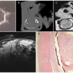
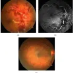
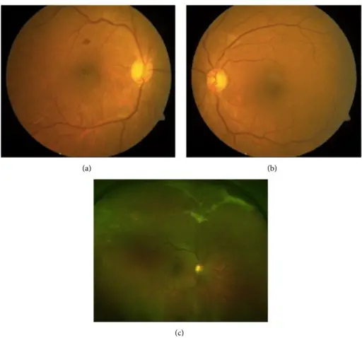
Sickle cell retinopathy is diagnosed by fluorescein angiography, spectral domain optical coherence tomography, and funduscopic examination.
How is Sickle Cell Retinopathy Treated?
Sickle cell retinopathy is treated with hyperbaric oxygen therapy, hydroxyurea, hydroxycarbamide treatment, intravitreal anti-vascular endothelial growth factor, scatter laser coagulation, and surgery.
What is the Prognosis of Sickle Cell Retinopathy?
The prognosis of sickle cell retinopathy is some do not survive beyond infancy, reduced life expectancy as symptoms manifest. New treatments are improving life expectancy.