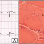
Changes of Purkinje cell morphology in cases with spinocerebellar ataxia type 31 (SCA31) (A and B: control subject; C–J: SCA31 case). (A) Purkinje cells of a control subject have round nuclei. (B) Thin neurites (arrows) around a Purkinje cell are seen in a control subject. (C) Two normal-sized Purkinje cells are shown in an SCA31 case. One has a dark cytoplasm and nucleus, suggesting that it has started to shrink (arrowhead). The other apparently has the halo-like amorphous materials with a bent nucleus (thin arrow). (D and E) Bright and elongated nuclei (thin arrows) are occasionally folded up at their end (D, thin arrow) in Purkinje cells with the halo-like amorphous materials. (F) Degenerated Purkinje cell with the typical halo-like amorphous materials. Purkinje cells are shrunken and their nuclei become severely deformed, rod-shaped, and condensed. (G) Ghost-like feature of a Purkinje cell is shown, where the outline of the nucleus is obscure. (H) Ghost-like feature of a Purkinje cell surrounded by bundles of lines (thin arrows). (I) The cell body of Purkinje cell is obscured in the empty basket formed by thick neurites (arrows). (J) A severely thin Purkinje cell with a shrunken and condensed nucleus that was not surrounded by the halo-like amorphous materials. A, C–H, and J: HE stain, B and I: Bodian stain. Bars; 20 μm. Distinctive features of degenerating Purkinje cells in spinocerebellar ataxia type 31. Yoshida K, Asakawa M, Suzuki-Kouyama E, Tabata K, Shintaku M, Ikeda S, Oyanagi K - Neuropathology : official journal of the Japanese Society of Neuropathology (2013). Not Altered. CC.
Spinocerebellar ataxia is a genetically heterogeneous group of inherited progressive degenerative disorders, the clinical hallmark of which is loss of balance and coordination accompanied by slurred speech.
What is the Pathology of Spinocerebellar Ataxia?
The pathology of spinocerebellar ataxia is:
-Etiology: The cause of spinocerebellar ataxia is mutations in many different genes cause the different types of spinocerebellar ataxia (SCA).
-Genes involved: ATXN1 gene mutations that cause SCA1 involve a DNA segment known as a CAG trinucleotide repeat.
-Pathogenesis: The sequence of events that lead to spinocerebellar ataxia are reduction in mass of frontal, temporal, and parietal portions of the brain along with the cerebellar peduncles, brainstem, and cranial nerve III.
How does Spinocerebellar Ataxia Present?
Patients with spinocerebellar ataxia are typically males. The symptoms, features, and clinical findings associated with Spinocerebellar ataxia include incoordination of gait and poor coordination of hands, speech, and eye movements, parkinsonism, chorea, pyramidalism, cognitive impairment, peripheral neuropathy, and seizures.
How is Spinocerebellar Ataxia Diagnosed?
Spinocerebellar ataxia is diagnosed with genetic testing.
How is Spinocerebellar Ataxia Treated?
Spinocerebellar ataxia has no cure. Zolpidem or varenicline may improve symptoms.
What is the Prognosis of Spinocerebellar Ataxia?
The prognosis of spinocerebellar ataxia is fair.


