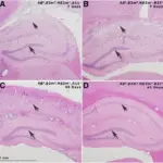
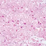
Subacute neuronal injury is neuronal death due to a progressive disease. The death is usually due to apoptosis and is associated with reactive gliosis. This includes cognitive defects, confusion, and impaired information processing.
What is Subacute Neuronal Injury?
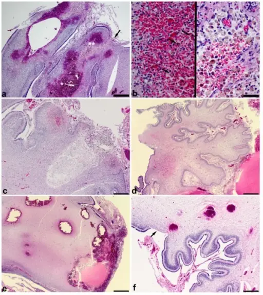
Histopathology of CHI demonstrates a multifocal hemorrhagic process and is associated with cortical hypoxic-ischemic injury or atrophy. a Multiple hemorrhages arising from different folia with multifocal extension into the leptomeninges in an infant of 25 weeks gestation who survived about 3 weeks. There are multiple areas of focal cortical loss, one of which is indicated by an arrow. Hemorrhages appear to be centered in the folia and show a mixture of acute and subacute changes suggesting that CHI may be due to repeated hemorrhagic episodes. b Left panel shows a higher magnification of the area marked by * in a demonstrating acute hemorrhage with scattered hemosiderin-laden macrophages (arrows). Right panel shows the area marked by ** in a at higher magnification demonstrating many hemosiderin-laden macrophages in an organizing cavity. c Chronic CHI with cavitation, hemosiderin and cholesterol clefts in the brain depicted in Figure 1e. d Atrophic cerebellar cortex uninvolved by hemorrhage from the sagittal section depicted in Figure 2c. e A larger hemorrhage extending into the leptomeninges with other smaller hemorrhages nearby in the white matter suggesting that the larger hemorrhages may represent a coalescence of multiple smaller hemorrhages (gross pathology is depicted in Figure 2a,b). f Three acute hemorrhages in the deep cortex and superficial white matter in a 29 weeks gestation infant who survived 3 weeks. The focal loss of gliotic cerebellar cortex (arrow) is consistent with a hypoxic-ischemic episode. All photomicrographs are taken from hematoxylin and eosin stained sections. a bar is 500 μm. b bar is 50 μm. c bar is 1 mm. d- f bar is 500 μm.Cerebellar hemorrhagic injury in premature infants occurs during a vulnerable developmental period and is associated with wider neuropathology.
Haines KM, Wang W, Pierson CR - Acta neuropathologica communications (2013). Not Altered. CC.
Post navigation
Previous Post
 What are Red Neurons?
Next Post
What is Chronic Neuronal Injury?