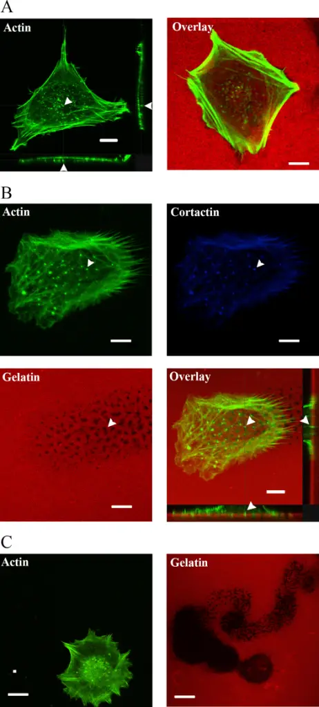
(A) Trophoblast cell stained with AlexaFluor-488-Phalloidin show actin rich structures on the ventral surface of the cell when plated on plastic. When plated on fluorescently labelled gelatin (0.2%, w/v), degradation of the extracellular matrix is observed. (B) Trophoblast cell displaying similar actin rich structures as in A which are also cortactin rich (blue), similar to the composition of invadosomes. Cells were also plated on fluorescently labelled gelatin and the overlaid image of actin (green) and gelatin (red) shows that these structures were able to invade and degrade the fluorescent matrix. (C) Trail of degradation by a migrating trophoblast cell through a fluorescently labelled matrix. Formation of atypical podosomes in extravillous trophoblasts regulates extracellular matrix degradation: Patel A, Dash PR - European journal of cell biology (2012). Not altered. CC.
The extracellular matrix are the components outside of the cell.
Cells interact with the extracellular matrix.
