Thromboembolism is the obstruction of a blood vessel by a blood clot. It is responsible for approximately 95% of embolisms.
What is the Pathology of Thromboembolism?
The pathology of thromboembolism is:
-Etiology: The cause of thromboembolism is family history, sedentary lifestyle, older age, obesity, smoking, high blood pressure, high cholesterol, and diabetes.
-Genes involved: Certain clotting factor abnormalites may be implicated.
-Pathogenesis: The sequence of events that lead to thromboembolism depends upon Virchow’s triad, i.e, presence of abnormalities of blood flow, blood vessel wall, and blood clotting components.
-Morphology: The morphology associated with thromboembolism shows large embolus may embed in major branches or lodge astride the bifurcation as a saddle embolus.
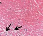
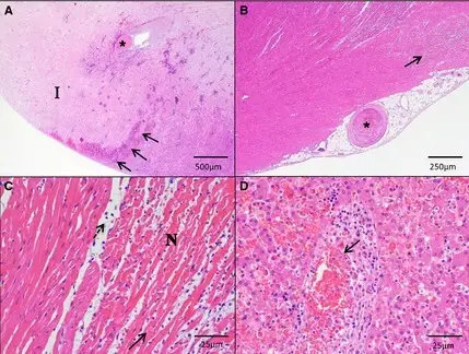
-Histology: The histology associated with thromboembolism shows necrosis, recanaliztion, irregular, nonlaminar, often obliterative, intimai fibrosis.
How does Thromboembolism Present?
Patients with thromboembolism are typically males as compared to females present at the age range of over 60. The symptoms, features, and clinical findings associated with thromboembolism include heart attack, limb infarction, or even pneumonia, leg pain and tenderness, edema, sense of impending doom, anxiety, red and hot skin, and dilated veins.
How is Thromboembolism Diagnosed?
Thromboembolism is diagnosed using D-dimer blood test, factor VIII blood test, platelet aggregation test, MRI, duplex ultrasound.
How is Thromboembolism Treated?
Thromboembolism is treated with anticoagulants, antiplatelets, and thrombolytics initially, if progressed then surgery is an option.
What is the Prognosis of Thromboembolism?
The prognosis of thromboembolism is fair, with short-term survival ranging from 95% to 97% for deep vein thrombosis, while long-term survival ranges from 61% to 75% for deep vein thrombosis.