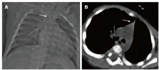
Thymic hypoplasia results in aberrant T-cell production, which negatively effects immune respnse. Thymic hypoplasia is also knowns as DiGeorge syndrome.
What is the Pathology of Thymic Hypoplasia?
The pathology of thymic hypoplasia aka DiGeorge syndrome is:
-Etiology: The cause of hypoplasia aka DiGeorge syndrome is a heterozygous deletion of part of the long arm (q) of chromosome 22, region 1, band 1, sub-band 2.
-Pathogenesis: The sequence of events that lead to thymic hypoplasia aka DiGeorge syndrome are abnormal development in the embryonic pharyngeal system. The T cell defects are the result of insufficient thymic tissue.
-Morphologic changes: The morphologic changes involved with thymic hypoplasia aka DiGeorge syndrome include: thymic hypoplasia, an abnormal chin, low set ears, and widely spaced apart eyes. Patients with thymic hypoplasia may also have cleft lips or cleft palates.
How does Thymic Hypoplasia Present?
Some signs and symptoms may be apparent at birth, but others may not appear until later in infancy or early childhood. The symptoms, features, and clinical findings associated with DiGeorge syndrome include: hearing and vision problems, mouth and feeding problems, delays in learning to walk or talk, short stature, frequent infections.
How is Thymic Hypoplasia Diagnosed?
Thymic hypoplasia or DiGeorge syndrome is diagnosed by fluorescent in situ hybridization (FISH) analysis which looks for the specific abnormal changes on chromosomes that cause the disease whcih is 22q11.2. Fluorescent in situ hybridization can be performed on cell samples obtained by amniocentesis as early as the fourteenth week of pregnancy. Diagnosis can be made by ultrasound examination around the eighteenth week of pregnancy, when abnormalities in the development of the heart or the palate can be detected.
How is Thymic Hypoplasia Treated?
Thymic hypoplasia aka DiGeorge syndrome is treated by a transplant of thymus tissue. This transplantation promotes T cell development. A cleft lip of cleft palate may be surgically repaired.
What is the Prognosis of Thymic Hypoplasia?
The prognosis of thymic hypoplasia aka DiGeorge syndrome depends on the severity of congenital disabilities. Some of these conditions may be life-threatening. With ongoing treatment and support, many individuals with DiGeorge syndrome may live active lives.


