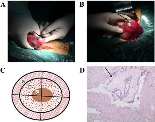
Lesion appearance, schema chart and pathological section. (A) Laparotomy revealed a mass inside the left posterior lateral aspect of the uterus. (B) Image of the mass removed. (C) Schema chart of the lesion section: a, normal muscle tissue in the outer layer; b, blood clot surrounding the vesicle; and c, edematous vesicle. (D) Pathological section demonstrating villi inside the uterine wall isolated from the uterine cavity, as demonstrated by the black arrow (hematoxylin and eosin stained; magnification, ×400). Distinguishing between intramural pregnancy and choriocarcinoma: A case report. Oncology Letters. Not Altered. CC.
Uterus pathology is the study and diagnosis of disease of uterus which is a hollow muscular organ located in the female pelvis between the bladder and rectum.
Examples of uterus pathology include:
- Functional endometrial disorders
- Inflammatory disorders of the endometrium
- Adenomyosis
- Endometriosis
- Endometrial polyps
- Endometrial hyperplasia
- Malignant tumors of the endometrium
- Tumors of endometrial stroma
- Tumors of the myometrium


