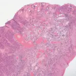
Vulvar intraepithelial neoplasia is a precancerous skin condition on the vulva. It happens when there are changes in the cells of the skin covering the vulva.
What is the Pathology of Vulvar Intraepithelial Neoplasia?
The pathology of vulvar intraepithelial neoplasia is:
-Etiology: The cause of vulvar intraepithelial neoplasia is not known exactly, but herpes simplex virus and human papilloma virus are linked with it.
-Genes involved: None.
-Pathogenesis: The sequence of events that lead to vulvar intraepithelial neoplasia involve viral DNA integration into the cells causing production of oncoproteins which affect normal cellular function, allowing neoplastic cells to evade apoptosis.
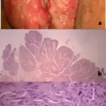
-Morphology: The morphology associated with vulvar intraepithelial neoplasia shows abnormal skin.
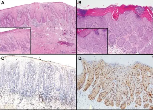
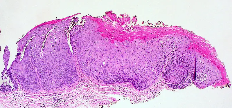
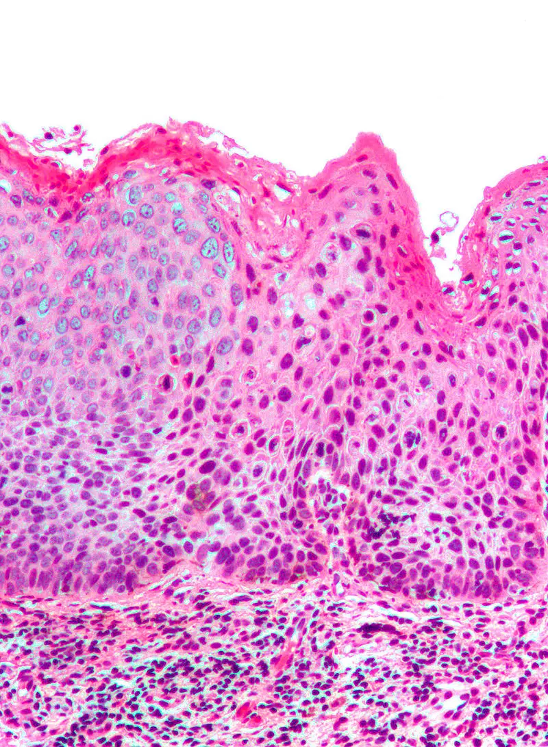
-Histology: The histology associated with vulvar intraepithelial neoplasia shows enlarged nuclei, hyperchromasia, and pleomorphic cells.
How does Vulvar Intraepithelial Neoplasia Present?
Patients with vulvar intraepithelial neoplasia are females, usually in their 40s. The symptoms, features, and clinical findings associated with vulvar intraepithelial neoplasia include tingling or burning in vulvar region, soreness, redness, and painful intercourse.
How is Vulvar Intraepithelial Neoplasia Diagnosed?
Vulvar intraepithelial neoplasia is diagnosed by physical exam, coloposcopy, and biopsy.
How is Vulvar Intraepithelial Neoplasia Treated?
Vulvar intraepithelial neoplasia is treated by excision of the affected area, laser ablation, or topical imiquimod treatment.
What is the Prognosis of Vulvar Intraepithelial Neoplasia?
The prognosis of vulvar intraepithelial neoplasia is good. Please note that if the condition is left untreated it may progress towards invasive carcinoma.

