Leiomyomas are benign tumors that originate in smooth muscle cells of the myometrium, which is the thick middle layer of the uterine wall.
What is the Pathology of Leiomyomas?
The pathology of leiomyomas is:
-Etiology: The cause of leiomyomas is genetic mutations in the smooth muscle cells.
-Genes involved: HMGA1 and HMGA2, RAD 51L1, PCPE gene.
-Pathogenesis: The sequence of events that lead to leiomyomas are not well understood. However, Genetic predisposition, environmental factors, steroid hormones, and growth factors involved in fibrotic processes and angiogenesis all play a role in the formation and growth of uterine fibroids hence forming the leiomyomas.
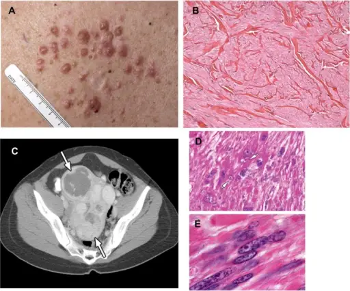
-Morphology: The morphology associated with leiomyomas shows dense, well-circumscribed nodules with white to tan colored cut surfaces.
-Histology: The histology associated with leiomyomas shows monoclonal neoplasms surrounded by a thin pseudocapsule of areolar tissue and compressed muscle fibers.
How does Leiomyomas Present?
Patients with leiomyomas typically females with increased incidence up to menopause. The symptoms, features, and clinical findings associated with leiomyomas include: Heavy menstrual bleeding, pelvic pain, periods lasting more than a week, infertility, abnormal bleeding, large masses, pain.
How is Leiomyomas Diagnosed?
Leiomyomas is diagnosed by comprehensive physical examination, ultrasound, and biopsy.
How is Leiomyomas Treated?
Leiomyomas is treated by surgical removal, hysterectomy, or myomectomy. Medications may be used for pain management.
What is the Prognosis of Leiomyomas?
The prognosis of leiomyomas is excellent. Intraperitoneal hemorrhage caused by uterine leiomyomas can be a life-threatening condition.


