Lung Cancer
In the United States of America (USA), lung cancer is the cancer that kills people most commonly.
Key risk factors for lung cancer include:
- Cigarette smoke
- Radon exposure
- Asbestos exposure
Because of the more than 60 carcinogens found in cigarette smoke, people who smoke account for 85% of lung cancer cases.
Particularly mutagenic substances in cigarettes include arsenic and polycyclic aromatic hydrocarbons.
Smoking duration and amount, or “pack-years,” are directly correlated with the chance of developing cancer.
The radioactive decay of uranium, which is found in soil, produces radon.
Uranium accumulates in enclosed areas like basements.
Uranium was identified to be the second most common cause of lung cancer in the US and is responsible for the majority of the public’s exposure to ionizing radiation.
Uranium miners also have a higher chance of developing lung cancer.
The non-specific symptoms of lung cancer include:
- Coughing
- Weight loss
- Hemoptysis
- Postobstructive pneumonia
The diagnosis of malignancy requires a biopsy even though imaging frequently identifies a single nodule or coin-lesion.
A “coin-lesion” can also be caused by benign lesions, which commonly affect younger patients.
Examples of lung lesions include:
- Granuloma
- Bronchial hamartoma
- Lung carcinoma
- Small cell carcinoma
- Non-small cell carcinoma
A small area of inflammation is called a granuloma.
In the Midwest, granulomas are frequently caused by histoplasma or tuberculosis.
Bronchial hamartomas are benign tumors composed of lung tissue and cartilage.
Lung carcinoma is a type of cancer that begins in the lungs.
Lung carcinoma is classically divided into two categories:
- Small cell carcinoma
- Non-small cell carcinoma
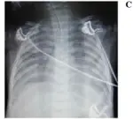
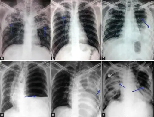
 Lung Cancer. Primary pulmonary sarcoma in an asymptomatic 72-year-old male Rosen . Not altered. CC BY-SA 2.0


Small Cell Lung Carcinoma
Small cell carcinoma, which comprises 15% of lung cancers, is an undifferentiated tumor made up of cells that look primitive.
Small cell carcinoma is usually not amenable to surgical resection but is treated with chemotherapy.
 Small Cell Lung Carcinoma. Histopathologic image of small-cell carcinoma of the lung. CT-guided core needle biopsy. No machine-readable author provided. KGH assumed (based on copyright claims). - Not altered. CC BY-SA 3.0


Non-Small Cell Lung Carcinoma
Non-small cell lung carcinoma is the majority of lung tumors.
Approximately 85% of lung tumors are non-small cell lung cancers.
Non-small cell lung carcinoma does not respond well to chemotherapy, thus is initially treated with surgical resection.
Subtypes of non-small cell lung carcinoma include:
- Adenocarcinoma (40%)
- Squamous cell carcinoma (30%)
- Large cell carcinoma (10%)
- Neuroendocrine tumor of the lung aka carcinoid tumor (5%)
TNM staging is the process that involves determining the extent and location of a person’s cancer.
Tumor size and local extension can be observed as well.
Adenocarcinoma is typically associated with pleural involvement.
Superior vena cava (SVC) obstruction results in enlarged head and neck veins, edema, and blue coloring of the arms and face called superior vena cava syndrome (SVCS).
Involvement of recurrent laryngeal (hoarseness) or phrenic (diaphragmatic paralysis) nerve.
Horner syndrome is caused by compression of the sympathetic chain.
Symptoms of Horner syndrome include:
- Ptosis
- Drooping eyelids
- Miosis (excessive constriction of the pupil)
- Pinpoint pupils
- Anhidrosis
Pancoast tumors may cause Horner syndrome.
The adrenal gland is a distinctive location for distant metastases.
Overall, the 5-year survival rate for non-small cell carcinoma illness is only 15%.
As a result of the lack of a reliable screening procedure, non-small cell carcinoma frequently manifests late.
 Non-Small Cell Lung Carcinoma. Pie chart showing incidences of nonsmall-cell lung cancers as compared to small-cell carcinoma shown at right, with fractions of smokers versus nonsmokers shown for each type. Mikael Häggström. Not altered. CC0


Adenocarcinoma of the Lung
Adenocarcinoma of the lung is composed of glandular cells.
If a non-smoker has lung cancer, it is likely adenocarcinoma of the lung (opposed to squamous cell carcinoma).
Adenocarcinoma of the lung tends to be peripherally located.
 Adenocarcinoma of lung, FNA. Ed Uthman. Not altered. CC BY 2.0
")


Squamous Cell Carcinoma of the Lung
Squamous cell carcinoma of the lung is typically centrally located.
Squamous cell carcinoma of the lung is more common in smokers.
 Squamous Cell Carcinoma of the Lung.The tumor involves the bronchus (probable site of origin) and extends deeply into lung parenchyma. Tumor nodules are present in the lower lung The pleura is focally involved. Yale Rosen. Not altered. CC BY-SA 2.0

Large Cell Carcinoma of the Lung
Large cell carcinoma of the lung typically grows quickly.
Large cell carcinoma of the lung spreads aggressively.
 Large Cell Carcinoma of the Lung. The corresponding surgical resection shows neoplastic cells with abundant pale eosinophilic cytoplasm and a surrounding infiltrate of inflammatory cells which can also be seen among the tumor cells in the fine needle aspirate specimen. A histologic section shows a proliferation of atypical cells along the alveolar walls (A). The Armed Forces Institute of Pathology (AFIP) - Not altered. Public Domain


Neuroendocrine Tumor of the Lung
Neuroendocrine tumor (NET) of the lung is composed of neuroendocrine cells.
Histology of neuroendocrine tumor of the lung shows cells with salt and pepper chromatin.
 Neuroendocrine Tumor of the Lung. Mitoses in a neuroendocrine tumor. Mikael Häggström, M.D. CC0