Vagina
The vagina is the canal that leads to the cervix.
The vagina is lined by non-keratinizing squamous epithelium.
Vagina pathology includes:
- Lichen sclerosus
- Extramammary Paget disease
- Adenosis
- Clear cell adenocarcinoma
- Embryonal rhabdomyosarcoma
- Vaginal carcinoma
- Vulvar carcinoma
 Vagina. Schematic drawing of female reproductive organs, frontal view. CDC, Mysid. Not altered. Public Domain
Lichen Sclerosis
Lichen sclerosis is a benign skin condition that often affects the female genital region.
Lichen sclerosis appears as wrinkly white thin skin.
Symptoms of lichen sclerosis include:
- Pain
- Irritation
- Itching
Lichen sclerosis is a risk factor for vulvar carcinoma.
 Lichen Sclerosis. Micrograph of extragenital lichen sclerosus: epidermal atrophy, follicular plugging and basal vacuolization, and sclerosis with initial homogenization of collagen in the dermis.[23] Anna Jędrowiak, Aleksandra Kobusiewicz, Ewa Trznadel-Grodzka, Andrzej Kaszuba - (2018). "Dermoscopic findings in extragenital lichen sclerosus". Our Dermatology Online 9 (2): 197–199. DOI:10. Not altered. CC BY 4.0

Extramammary Paget Disease
Extramammary Paget disease is characterized by the presence of malignant epithelial cells in the vulva’s epidermis.
Symptoms of extramammary Paget disease include erythematous, itchy, and ulcerated vulvar skin.
Extramammary Paget disease represents a carcinoma in situ, which is typically a surface cancer.
 Extramammary Paget Disease. H&E stained micrograph of Extramammary Paget's disease, showing Paget cells infiltrating the epidermis Nephron. Not altered. CC BY-SA 3.0


Adenosis
Adenosis is due to concentrated columnar epithelium persistence in the upper third of the vagina.
During development:
- The columnar epithelial lining of the upper 1/3 of the vagina is derived from the Mullerian ducts
- The squamous epithelium from the lower 2/3 of the vagina derived from the urogenital sinus
There is increased occurrence of adenosis in females exposed to diethylstilbestrol (DES) while they were pregnant.
 Adenosis. Histopathology of sclerosing adenosis. Mikael Häggström, M.D Not altered. CC0
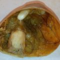
Clear Cell Adenocarcinoma
Clear cell adenocarcinoma is a malignant proliferation of glands with clear cytoplasm.
Clear cell adenocarcinoma is a complication of DES-related vaginal adenosis.
The use of diethylstilbestrol (DES) was discontinued as a result of the discovery of this consequence and other DES-related abnormalities of the gynecologic tract, such as irregular uterine shape.
 Ataxia-Telangiectasia. Hematoxylin and eosin stained sections of the gastric adenocarcinoma resected from our patient. [A] Cytological features of malignant glands; the cells are irregularly shaped with high nucleus to cytoplasm ratio and loss of nuclear polarity. The small dark cells are inflammatory cells (100× enlargement) [B] This area of tumor is in the serosa; there is redemonstration of irregular glands formed by tumor cells of varying sizes and orientation, with prominent nucleoli. The large clear spaces are fat cells (200× enlargement). Gastric outlet obstruction due to adenocarcinoma in a patient with Ataxia-Telangiectasia syndrome: a case report and review of the literature. Otabor IA, Abdessalam SF, Erdman SH, Hammond S, Besner GE - World journal of surgical oncology (2009). Not Altered. CC.
Embryonal Rhabdomyosarcoma
Embryonal rhabdomyosarcoma is a malignant mesenchymal growth involving malignant muscle cells.
Sarcoma botryoides is a specific type of embryonal rhabdomyosarcoma.
Sarcoma botryoides presents as a bleeding grape-like mass that protrudes from the vagina.
The histology of sarcoma botryoides has a distinctive cell, the rhabdomyoblast, that has cytoplasmic cross-striations and is immunohistochemically stained positively for desmin and myogenin.
 Embryonal Rhabdomyosarcoma. Embryonal rhabdomyosarcoma consisting almost entirely of differentiated rhabdomyoblasts (H&E staining, ×40). Embryonal rhabdomyosarcoma of upper lid in 15-year-old patient. Sharifi M - Case reports in ophthalmological medicine (2014). Not Altered. CC.

Vaginal Carcinoma
The squamous epithelium that lines the vaginal mucosa can develop into cancer.
Vaginal carcinoma is typically associated with high-risk HPV.
Vaginal intraepithelial neoplasia (VIN) is the precursor lesion of vaginal carcinoma.
When local lymph nodes are affected, cancer from the bottom two-thirds of the vagina spreads.
Vaginal carcinoma from the lower third travels to the regional iliac nodes, and cancer from the inguinal nodes.
 Vaginal Carcinoma. Micrograph of a mucinous adenocarcinoma Nephron. Not altered. CC BY-SA 3.0
Vulvar Carcinoma
Multiple partners and an early age of sexual contact are risk factors connected to HPV exposure, which typically affects women of reproductive age.
Vulvar carcinoma arises from vulvar intraepithelial neoplasia (VIN).
Vulvar intraepithelial neoplasia is a dysplastic precursor lesion to vulvar carcinoma with:
- Abnormal nuclear atypia
- Koilocytic alteration
- Enhanced mitotic activity
The most frequent cause of non-HPV associated vulvar cancer is chronic lichen sclerosus.
Chronic inflammation and irritation subsequently result in carcinoma.
Vulvar carcinoma is typically observed in older women (mean age > 70-years-old).
 Vulvar Carcinoma. Stage 4A vulvar cancer Cancer Research UK - Not altered. CC BY-SA 4.0